Early Heart Risk Detection
Routine Eye Scans May Detect Atrial Fibrillation Years Before Formal Diagnosis
Anonymous
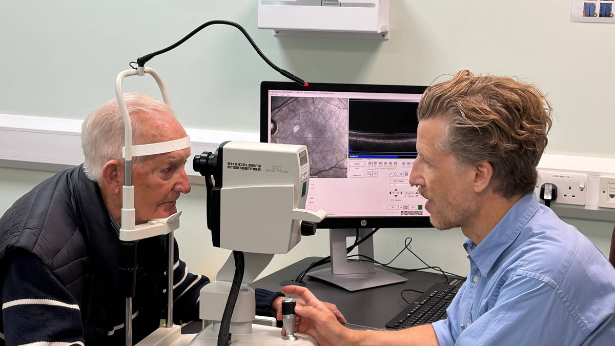
Researchers found that retinal imaging may help identify atrial fibrillation years before diagnosis, potentially enabling earlier treatment to reduce the risk of stroke and heart failure. An analysis of more than 90,000 people found that patients with atrial fibrillation—and those who later developed it—had thinner layers of retinal nerve cells, with differences detectable up to four years before diagnosis. Because atrial fibrillation can be intermittent and missed by standard electrocardiograms, researchers say routine eye scans could offer a noninvasive screening tool, though further validation is needed. Separately, a Swedish trial found that adding targeted treatment of scarred, low-voltage areas in the heart to standard catheter ablation substantially reduced recurrence of persistent atrial fibrillation: 67.6% of patients remained free of arrhythmias after 12 months, compared with 37.4% receiving standard treatment alone.
Key Points
1
The retinal analysis combined two distinct datasets and imaging methods: optical coherence tomography scans and color retinal photographs from the AlzEye cohort at Moorfields, which included more than 50,000 patients, and the UK Biobank cohort of roughly 40,000 participants. Of those cohorts, 5,735 and 526 people, respectively, already had atrial fibrillation.
2
The retinal biomarker was specifically a thinner macular ganglion cell and inner plexiform layer (mGCIPL), a nerve-cell layer that connects the eye to the brain; the study found that greater thinning was associated with a higher likelihood of atrial fibrillation.
3
Because retinal imaging is already routinely performed in community optometry practices and hospital eye clinics, researchers said it could potentially be incorporated into existing eye-care pathways rather than requiring a wholly new screening infrastructure.
4
The ablation trial involved 209 patients with persistent atrial fibrillation and clearly defined low-voltage zones in the left atrium across five Swedish hospitals. Patients were randomly assigned to pulmonary-vein isolation alone or pulmonary-vein isolation plus targeted treatment of those low-voltage areas, with outcomes assessed after 12 months.
5
The Swedish researchers focused on low-voltage zones because they indicate fibrosis or scarring, where electrical signals are weaker than in healthy heart tissue; the added procedure was intended to interrupt arrhythmia-related electrical activity in these diseased areas.
Sentiment
Mostly NegativeNEGATIVENEUTRALPOSITIVE
Publishers
8
Articles
4
Reach
12
Topics
A
AnonymousAuthor


